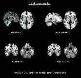
Brain Volume Alterations in SUDEP
Abstract number :
2.170
Submission category :
5. Neuro Imaging / 5A. Structural Imaging
Year :
2018
Submission ID :
501352
Source :
www.aesnet.org
Presentation date :
12/2/2018 4:04:48 PM
Published date :
Nov 5, 2018, 18:00 PM
Authors :
Luke Allen, University College London; Sjoerd B. Vos, Centre for Medical Image Computing, University College London; Epilepsy Society, Chalfont St Peter; Ronald Harper, UCLA Brain Research Institute; Rajesh Kumar, UCLA Brain Research Institute; Jennifer A
Rationale: Structural neuroimaging studies have outlined volumetric alterations among important autonomic and respiratory regulatory brain structures in sudden unexpected death in epilepsy (SUDEP) and those at greatest risk (1,2,3). However, neuroimaging studies of SUDEP have typically involved small sample sizes. Furthermore, whole-brain deformation-based studies, which allow exploration of white and gray matter changes, remain lacking. Here, we analysed whole-brain volumes in 28 SUDEP cases, age- and sex-matched healthy controls (n=28), as well as living age-, sex- and epilepsy syndrome-matched high-(n=28) and low-risk (n=28) epilepsy controls. Methods: We used voxel-based morphometry (VBM) and deformation-based morphometry (DBM) procedures to assess gray and white matter in SUDEP, and high- and low-risk patients compared with healthy subjects. Image analysis was performed using the computational anatomy toolbox (3) within SPM12 (4). P-values were false discovery rate (FDR) corrected. Results: VBM showed gray matter volume loss in SUDEP and patients at high-risk compared with healthy controls, with the posterior-cingulate, left thalamus, dorsal midbrain/periaqueductal gray and cerebellar cortex most affected in SUDEP. Right amygdala gray matter volume was increased in SUDEP and high-risk patients. DBM showed extensive volume reductions in the gray and white matter of the limbic system, deep “autonomic” nuclei and cortex of the cerebellum, and temporal and frontal lobes in SUDEP and to a similar extent, in high-risk patients, but to a much lesser extent in low-risk subjects. Conclusions: SUDEP is associated with volume loss in the limbic system (particularly in the hippocampus and cingulate), the lateral thalamus, cerebellum, and dorsal midbrain/periaqueductal gray of the brainstem. Patients at high-risk (those having frequent generalised tonic clonic seizures) show similar patterns of volume alteration. Loss or injury to neurons in these regions, particularly in cerebellar sites that dampen extremes of blood pressure change or assist in recovery from apnea, as well as suprapontine sites known to trigger or inhibit inspiratory efforts pose a risk for SUDEP. The graded nature of these imaging features with risk suggests the potential to non-invasively and prospectively identify patients at greatest peril of a fatal outcome. Funding: This work was undertaken at UCLH/UCL who receives a proportion of funding from the Department of Health’s NIHR Biomedical Research Centres funding scheme. This work was funded by the NIH – National Institute of Neurological Disorders and Stroke U01-NS090407-01 (The Center for SUDEP Research). Functional MRI data acquired through Medical Research Council funding (grant number G0301067). SBV is funded by the National Institute for Health Research University College London Hospitals Biomedical Research Centre (NIHR BRC UCLH/UCL High Impact Initiative). We are grateful to the Wolfson Foundation and the Epilepsy Society for supporting the Epilepsy Society MRI scanner.

.tmb-.png?Culture=en&sfvrsn=b4f4c966_0)