Electrocorticography and Surgical Outcomes in Intractable Epilepsy due to Temporal Encephalocele
Abstract number :
2.335
Submission category :
9. Surgery / 9C. All Ages
Year :
2018
Submission ID :
503464
Source :
www.aesnet.org
Presentation date :
12/2/2018 4:04:48 PM
Published date :
Nov 5, 2018, 18:00 PM
Authors :
Kiran Kanth, Mayo Clinic; Gregory D. Cascino, Mayo Clinic; Jeffrey W. Britton, Mayo Clinic; Elson So, Mayo Clinic; Karl Krecke, Mayo Clinic; Robert J. Witte, Mayo Clinic; Jamie J. Van Gompel; and Lily Wong-Kisiel, Mayo Clinic
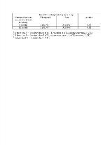
Rationale: Temporal encephalocele (TE) is an under-recognized surgically-remediable cause of medically refractory temporal epilepsy. The utility of intraoperative electrocorticography (iECog) and extraoperative electrocorticography (eECog) in guiding extent of surgical resection is unknown. This study assessed interictal discharges from iECog and eECog and surgical outcomes in patients with medically refractory temporal lobe epilepsy associated with TE. Methods: Patients with TE and medically refractory temporal lobe epilepsy who underwent surgical intervention at the Mayo Clinic Rochester between January 2008 and July 2017 were identified. Patients with a minimum of 3 month postoperative follow-up were included. For each patient, interictal discharges from iECog and eECog were classified as widespread if present in both hippocampal and lateral temporal lobe contacts and focal if interictal discharges were present only within the contacts around the immediate area of resection. Results: Fifteen patients were identified (female 80%, median age 44.5 years, interquartile range (IQR) 22.9-54.9 years). TE was identified in 7 patients preoperatively (left 2, right 3, bilateral 2), from post-operative radiology re-review in 7 (left 1, right 1, bilateral 5), and 1 intraoperatively (right). Thirteen patients were evaluated with iECog, and 2 patients underwent eECog, 1 with grids, strips, and depth electrodes and 1 with stereotactic electroencephalogram. Interictal discharges were widespread in mesial and neocortical temporal lobes in 12 of 15 patients. Three patients (20%) underwent focal TE or limited temporal pole resection, 11 patients (73.3%) anterior temporal lobectomy (ATL), and 1 patient amygdalohippocampal laser ablation. There was no significant difference in seizure-free outcomes at 6 months or at 12 months between patients with widespread or focal interictal discharges from ECog (Table). Seven patients (46.7%) were seizure-free at last follow-up (median duration of follow-up 12.3 months, IQR 6.2-61.1 months). Seizure-freedom at last follow-up was seen in 1 of 3 patients with focal TE resection and 6 of 11 patients with ATL (p = 0.40). Focal TE resection was performed only in recent years (median duration of follow-up in focal resection 7 months, IQR 3.7-18.7, versus ATL 39.2 months, IQR 7.1-68.3), and comparison of surgical outcome between surgical groups was limited by to short-term follow-up. Conclusions: ECog often showed epileptogenic discharges extending beyond regions around the TE. Although interictal discharges on ECog might be used to guide surgical resection, interictal findings from iECog and eECog should be interpreted with caution given the limited follow-up of the recent approach of focal TE resection. Long-term follow-up is needed to clarify which surgical approach optimizes seizure outcome while limiting surgical resection. Funding: None
