EVIDENCE FOR PARIETO-FRONTAL DYSCONNECTION IN EPILEPTIC SYNDROMES WITH CONTINUOUS SPIKES AND WAVES DURING SLOW SLEEP
Abstract number :
A.07
Submission category :
Year :
2003
Submission ID :
4033
Source :
www.aesnet.org
Presentation date :
12/6/2003 12:00:00 AM
Published date :
Dec 1, 2003, 06:00 AM
Authors :
Xavier De Tiege, Serge Goldman, Steven Laureys, Denis Verheulpen, Catherine Chiron, Olivier Dulac, Patrick Van Bogaert PET/Biomedical Cyclotron Unit, ULB-Hopital Erasme, Brussels, Belgium; Department of Pediatric Neurology, ULB-Hopital Erasme, Brussels, B
Syndromes with continuous spikes and waves during slow sleep (CSWS) are age-related epileptic syndromes which associate cognitive dysfunctions and specific EEG abnormalities. Previous positron emission tomography (PET) studies using [18F] -fluorodeoxyglucose (FDG) have shown that some affected patients present focal hypermetabolism. We hypothesized that particular metabolic patterns could characterize this subgroup of patients.
Among epileptic children investigated by FDG-PET in our center, 11 patients, aged 3 to 10 years, presented the EEG pattern of CSWS and hypermetabolism on PET. These patients were considered as one group which was analyzed using a voxel-based method, Statistical Parametric Mapping 99, with a control group of young adults. In a first step, a simple comparison between the 2 groups was performed. In a second step, we searched for disease-induced changes in the contribution of a brain area to the level of metabolic activity in another brain area using [quot]pathophysiological interactions[quot] here defined as an application of the [quot]psychophysiological interactions[quot] to a pathological condition.
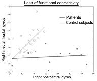
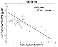
When compared with the control group, the patients group showed the coexistence of highly significant hypermetabolic areas in the right parietal lobe and hypometabolic areas in the frontal lobes ([italic]P[/italic][lt]0.05). Epilepsy-induced changes in the parieto-frontal metabolic relationships were then searched for. When considering the peak voxel values in the right parietal hypermetabolic areas, significant pathophysiological interactions with the metabolism in the right and the left frontal lobes were found ([italic]P[/italic][lt]0.05). Regression plots also indicated that hypermetabolic areas in the right parietal lobe were associated with a loss in functional connectivity with the right frontal lobe (figure 1) and inhibition in the left frontal lobe (figure 2).
This study brings evidences for parieto-frontal dysconnection in epileptic syndromes with CSWS. This altered modulation seems to be the consequence of a loss of functional connectivity or an inhibition mechanism. The presence of frontal hypometabolism is in agreement with the neuropsychological deficits frequently observed in epileptic patients with CSWS.[figure1][figure2]

