HIGH-ENERGY PHOPHORUS METABOLITES IN TEMPORAL LOBE EPILEPSY WITH HIPPOCAMPAL SCLEROSIS
Abstract number :
1.100
Submission category :
Year :
2005
Submission ID :
5151
Source :
www.aesnet.org
Presentation date :
12/3/2005 12:00:00 AM
Published date :
Dec 2, 2005, 06:00 AM
Authors :
R. Mark Wellard, Regula S. Briellmann, Matthew Litgermoet, Gaby S. Pell, and Graeme D. Jackson
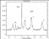
Hippocampal sclerosis (HS) is a small focal epileptogenic lesion. However, widespread subtle morphological abnormalities have been found in temporal lobe epilepsy with HS. Previous magnetic resonance spectroscopy (MRS) studies have suggested alterations in high-energy metabolites in temporal lobe of these patients. Here we assess both frontal and temporal lobes for abnormalities in high-energy metabolites, using phosphorus magnetic resonance spectroscopic shift imaging (31P-MRSI). We studied 15 healthy controls and 15 age-and gender matched TLE patients with unilateral HS. 31P-MRSI was recorded from two 3cm thick axial slices (temporal and frontal lobe), using a 10 x 10 phase encoding matrix over a 26 cm FOV. Regions of interest were the right and left temporal, and the right and left frontal lobe, 10-15 voxels were averaged for each ROI. Results for phosphocreatine (PCr); inorganic phosphate (Pi); beta-nucleoside triphosphate (ATP[beta]); and pH are given. These matabolites are labeled on the figure of a control spectra. Controls showed no difference between the right and left temporal or frontal lobe metabolites, therefore the mean of the two sides was used for comparison with patient data. The ratio PCr/Pi showed no difference between patients and controls, or between the ipsilateral and contralateral side of patients in both assessed lobes. The PCr/ATP[beta] ratio was higher in controls (0.88 [plusmn]0.1) than in the patients[apos] ipsilateral (0.63 [plusmn]0.2; p=0.001), or contralateral temporal lobes (0.76 [plusmn]0.07; p=0.05). The frontal lobe PCr/ATP[beta] ratio was not different between patients and controls. The pH was lower in controls (6.96 [plusmn]0.1) than in the patient[apos]s ipsilateral temporal lobe (7.01 [plusmn]0.1; p=0.04). There was no difference between controls and the patient[apos]s contralateral temporal lobe. The frontal lobe pH was not different between patients and controls. High-energy metabolites in TLE-HS patients show abnormalities in the region of the seizure focus, but also in the contralateral temporal lobes, but appear undisturbed in the frontal lobes. The reduction in the PCr/ATP[beta] ratio may reflect a higher energy demand, associated with the focal epileptic process.[figure1] (Supported by Neuroscience Victoria, the NHMRC Australia, the Brain Imaging Research Foundation and thank all the subjects for participation in our study.)
