MEASUREMENT OF TISSUE METABOLITES IN MRI-NORMAL TEMPORAL LOBE EPILEPSY PATIENTS
Abstract number :
2.216
Submission category :
Year :
2003
Submission ID :
2085
Source :
www.aesnet.org
Presentation date :
12/6/2003 12:00:00 AM
Published date :
Dec 1, 2003, 06:00 AM
Authors :
Todd K. Stevens, Warren T. Blume, Donald H. Lee, Samuel Wiebe, Seyed Mirsattari, David Diosy, Robert Bartha Department of Medical Biophysics, University of Western Ontario, London, ON, Canada; Department of Clinical Neurological Sciences, London Health Sc
Proton magnetic resonance spectroscopy (1H MRS) has become a valuable tool in detecting biochemical imbalances in the human brain. Short echo time (TE) 1H MRS can be used to measure tissue metabolites such as the excitatory neurotransmitters glutamate (Glu) and glutamine (Gln), N-acetylaspartate (NAA, marker for neuronal integrity), creatine (Cr, involved in cellular energy metabolism) and myo-inositol (Myo, a putative marker for gliosis). The purpose of this study is to determine metabolic abnormalities that are specific to the epileptogenic hippocampi of MRI-normal temporal lobe epilepsy (TLE) patients.
LASER localized (J Magn Reson. 2001; 153(2): 155-77) short TE 1H MRS data (TR/TE=3200/46 msec) were collected from 3 MRI normal bilateral TLE patients (2 males, 1 female, aged 37.3 [plusmn] 3.5 years, mean [plusmn] SD) using a quadrature hybrid birdcage radio frequency (RF) coil on a 4.0 Tesla Varian/Siemens Unity Inova whole body MRI. Adavanced spectral analysis techniques incorporating macromolecule subtraction, lineshape correction, and metabolite template modelling were employed to uniquely quantify Glu, Gln, NAA, Cr, Myo and 14 other metabolites from voxels in the left and right hippocampi (size = 2.70 [plusmn] 0.29 cm3). The water concentration of brain tissue was used as a reference to determine absolute metabolic concentrations, which were compared to levels found in the hippocampi of healthy controls (n=8, Magn Reson Med. 2003; 49(5): 918-27).
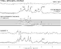
The residuals in Fig.1 demonstrate the consistency of the macromolecule removal and quality of spectral fitting from the spectra of two TLE patients. A preliminary multivariate analysis showed no overall group difference between the metabolite levels found in the epileptogenic hippocampi of the TLE patients and the control metabolite levels. However, one subject showed a significant bilateral reduction in NAA (Subject 1: Fig.1, [NAA] = 2.94 [plusmn] 0.50 mmol / L VOI) when compared with both controls ([NAA] = 9.18 [plusmn] 2.13 mmol / L VOI) and the other 2 TLE patients ([NAA] = 8.06 [plusmn] 0.65 mmol / L VOI).
Although preliminary, the results suggest that some MRI-normal TLE patients may have altered levels of NAA. Further studies are required to determine if other metabolites are also affected.[figure1]
[Supported by: Canadian Institiutes of Health Research (MME 15594), Robarts Research Institute.]
