Measuring seizure propagation speeds in humans with ictal high frequency activity
Abstract number :
3.295
Submission category :
Late Breakers
Year :
2013
Submission ID :
1851281
Source :
www.aesnet.org
Presentation date :
12/7/2013 12:00:00 AM
Published date :
Dec 5, 2013, 06:00 AM
Authors :
R. Connors, S. Weiss, G. Banks, G. McKhann, R. Goodman, R. Emerson, A. Trevelyan, C. Schevon
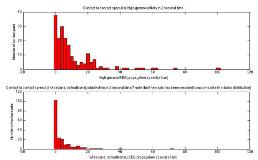
Rationale: Recordings of multi-unit activity during spontaneous human seizures have clearly distinguished between neuronal territories fully recruited into the seizure (characterized by high level and synchronized neuronal firing) and territories on the periphery of this core recruited area (characterized by low level and desynchronized firing). We use the terms ictal core and ictal penumbra for these two regions. Using multi-unit activity as a gold standard, we recently proposed ictal high frequency activity recorded with subdural macroelectrodes as a proxy of the ictal core. We hypothesized that the propagation speed of the ictal core, as defined by the spread of ictal high frequency activity, would be significantly slower than that of the wideband, or traditional, intracranial EEG (iEEG) ictal rhythm and would fall into a relatively narrow range of values across cortical pathologies. Methods: We performed a retrospective comparison of seizure propagation speeds in 18 seizures recorded in 8 patients. Pathology reports were available for all patients and included mesial temporal sclerosis, mesial temporal sclerosis in association with a cavernous angioma, non-specific right frontal gliosis, neocortical temporal malignant glioma, insular cortical dysplasia, and chronic neocortical infarction. We identified ictal high frequency activity by displaying traditional, unfiltered, intracranial EEG (iEEG) tracings adjacent to 80 to 150 band-pass filtered tracings. Channels were considered recruited into a seizure (ictal core) if they demonstrated an iEEG ictal rhythm in which greater than 50% of the wideband ictal waveforms were associated with high frequency activity at least 3 times the amplitude of the pre-ictal baseline. To address the problem of spurious oscillations in high pass filtered data raw iEEG data was inspected for the presence of rippling concomitant with high frequency activity; when uncertainty remained after examination of the raw iEEG data wavelet transformations were examined. Once all recruited channels were identified, each channel was reviewed individually and the earliest changes from the pre-ictal baseline in the traditional and 80-150 Hz band-pass filtered tracings were defined as the respective onset times. The absolute difference between all adjacent recruited channels (10 mm spacing) was defined as the ictal propagation speed. Results: Across all patients and seizures, the mean propagation speed of ictal high frequency activity, 0.94 mm s-1 +/- 0.78 mm s-1, was significantly faster than the mean propagation spread of the traditional ictal rhythm, 2.35 mm s-1 +/- 1.26 mm s-1 (p < 0.01, Welch s t-test). There was no significant difference in the propagations speeds of high frequency activity across separate pathological substrates (p = 0.20, one-way ANOVA). Conclusions: This work supports our hypothesis that the ictal core, as measured by ictal high frequency activity, propagates significantly more slowly than the traditional iEEG ictal rhythm and does so at a relatively narrow range of values across various pathologies.
