METABOLIC STATE CORRELATES WITH EXTRACELLULAR GLUTAMATE AND GABA IN TLE
Abstract number :
2.009
Submission category :
Year :
2005
Submission ID :
5313
Source :
www.aesnet.org
Presentation date :
12/3/2005 12:00:00 AM
Published date :
Dec 2, 2005, 06:00 AM
Authors :
1Idil Cavus, 2Kenneth Vives, 3Hoby P. Hetherington, 1John Krystal, 2Dennis D. Spencer, and 4Jullie W. Pan
Much imaging literature (PET and MR) and recent ex vivo studies have increasingly argued for a pathophysiological role of energy metabolism and mitochondrial function in TLE. However, it is difficult to directly demonstrate a link between dysfunctional energetics with in vivo hyperexcitability. The use of microdialysis catheters in human TLE patients provides an opportunity to evaluate the extracellular milieu in the seizure focus, and with imaging, to determine how extracellular (ECF) measures of glutamate and GABA may relate to metabolic imaging measures. N=7 unilateral hippocampal epilepsy patients (age, 33[plusmn]12years) were studied. These patients were studied pre-operatively using 1H and 31P spectroscopic imaging. 1H hippocampal data was collected using a modified LASER sequence with 0.64cc voxel resolution, angulating the plane along the temporal pole. 31P spectroscopic imaging was performed using a pulse acquire acquisition with 12cc voxel resolution.
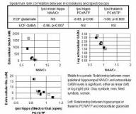
Microdialysis: Probes were attached to depth electrodes, implanted stereotaxically in regions of interest (MRI verified). To minimize post surgical effects, the catheters were not used until 2-5days after placement. The inter-ictal zero-flow study was performed at least 6hrs from any intracranially recorded seizure activity, and [gt]=2 hrs after any food intake. 20ul dialysate samples were collected at progressively decreasing flow rates and the samples were stored at [ndash]80[deg]C for later HPLC analysis. The microdialysis studies were typically performed within 3months of MR imaging. Because of the non-normal distribution of microdialysis data, rank correlations were used. Significant correlations were found for both extracellular glutamate and GABA (Figure). Notably with these data, no correlation was found between GABA and high energy phosphates, nor ECF glutamate and NAA/Cr. In this preliminary study, we detected correlations between metabolic imaging and extracellular neurotransmitter levels. That extracellular ipsilateral glutamate relates to PCr/ATP suggests that control of ECF glutamate reflects immediate available tissue energetics. The negative relationship of NAA/Cr with GABA may be consistent with a view that extracellular GABA is sensitive to mitochondrial capacity, wherein the elevation in GABA represents a response to deteriorating mitochondrial function.[figure1] (Supported by NIH RO1-NS40550 and PO1-NS39092.)
