Ripples (80-200 Hz) in the Human Mesial-Temporal Lobe Exhibit Different Profiles of Phase-Amplitude Coupling in Relation to Slow Waves
Abstract number :
1.032
Submission category :
1. Basic Mechanisms / 1C. Electrophysiology/High frequency oscillations
Year :
2018
Submission ID :
499197
Source :
www.aesnet.org
Presentation date :
12/1/2018 6:00:00 PM
Published date :
Nov 5, 2018, 18:00 PM
Authors :
Shennan Weiss, Thomas Jefferson University; Inkyung Song, Thomas Jefferson University; Sitaram Vangala, University of California - Los Angeles; Zachary Waldman, Thomas Jefferson University; Mustafa Donmez, Thomas Jefferson University; Itzhak Fried, David
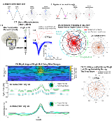
Rationale: Ripple oscillations (80-200 Hz) recorded from clinical electrodes in the mesial-temporal lobe (MTL) during slow wave sleep may be generated by physiological or pathological networks in patients with epilepsy. The mechanisms that distinguish physiological from pathological ripples are not yet clear. Methods: Intracranial EEG (iEEG)was recorded from 23 patients with mesial-temporal lobe epilepsy, and 17 patients with neocortical epilepsy. Also, iEEG with synchronized local field potentials (LFPs) were recorded from hybrid micro- and macroelectrodes implanted in the mesial-temporal lobe of five distinct patients during sleep. The microwire tips were 4mm away from the most distal iEEG contact. A custom pipeline was used to identify and quantify ripple oscillations in the iEEG as well as quantify phase-amplitude coupling relationships with the slow waves (0.1-2 Hz). Synchronized single unit activity was isolated from the LFP. Results: We found a higher probability of pathological MTL ripples (when MTL was within the SOZ) occurring around slow wave ‘peak-trough’ phases, corresponding to DOWN-UP transitions in two cohorts on patients. We established statistically in the first cohort (N=40, n=399 seizure onset zone [SOZ], n=708 non-SOZ) that a ripple’s preferred phase angle of coupling with respect to the slow wave depended on whether the electrode was within the SOZ (parametric two-way ANOVA for circular data, p<0.001, n=49,571), and not the power of the ripple event. In the five patients with hybrid electrode recordings we identified 60 excitatory single units, and two inhibitory single units from 19 mesial-temporal electrode sites. We detected a total of 10,407 slow wave coupled ripples above the second quartile of ripple power during 9 hours of iEEG sleep recording. Single units exhibited an increase in firing rate during ripple events associated with high power (above the second quartile). Slow wave coupled ripple events and synchronized single unit activity were categorized as ‘Trough to Peak’ or ‘Peak to Trough’ depending on the ripple’s preferred phase angle. The mean baseline single unit activity corresponding with the ripple events in the ‘Trough to Peak’ ripple distribution was greater than that of the ‘Peak to Trough’ distribution, indicating that the ‘Trough to Peak’ distribution included more of the UP state, while the ‘Peak to Trough’ distribution included more of the DOWN-UP transition. Ripples during the UP state were associated with a comparatively larger increase in single unit firing rates. Conclusions: In the epileptogenic mesial-temporal lobe pathological ripples occur during the DOWN-UP transition, while physiological ripples, that may be associated with memory consolidation, occur more often during the UP state. Distant from the ripple generator, a larger increase in single unit firing rates is seen during physiological ripples. Funding: SW is supported by NS094633. This work was also supported by NS033310 to J.E, and NS033221 to I.F.
